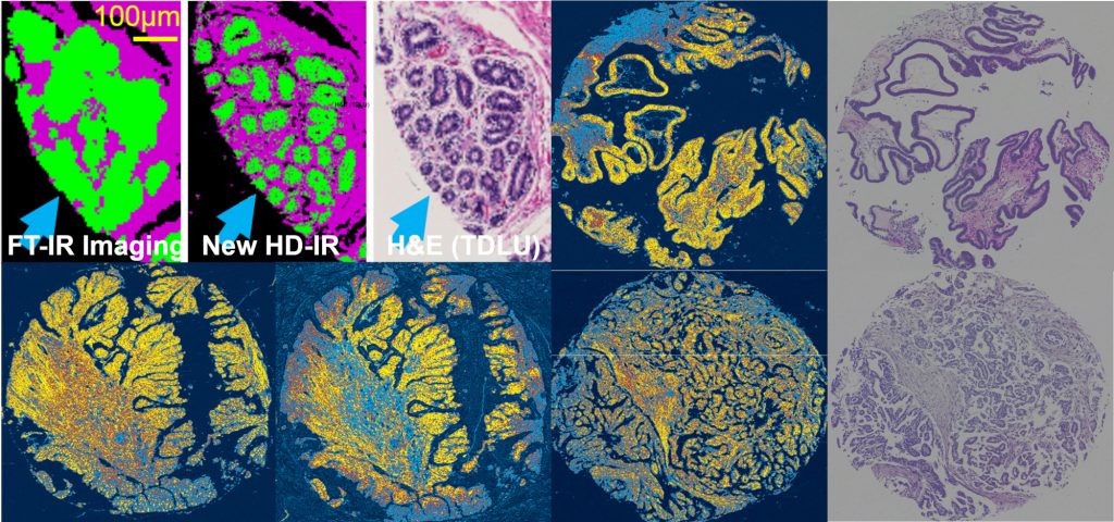
Fundamental molecular vibrational modes of organic molecules occur in the mid-infrared region of the electromagnetic spectrum. These vibrational modes result in sharp (resonance) peaks in the mid-infrared and have been used to identify molecules in homogeneous bulk materials for more than four decades. The chemical significance of peaks in the “fingerprint region” of the mid-infrared spectrum has also been tabulated extensively in spectroscopy literature. The chemical composition of substances can be ascertained by analyzing their mid-infrared spectrum. Combining spectroscopy with microscopy has been a relatively recent development and this has resulted in spatially resolved spectroscopic information. With this innovation, it is possible to obtain an image of a chemically diverse sample and discover the chemical composition at every pixel in that image. This chemical information has been used in a variety of fields from forensics to art restoration to performing completely automated histopathology in tissue sections. In tissue, the molecular composition and chemical detail at every pixel can be mapped over the entire imaging area and tissue function can be studied.
Mid-infrared spectroscopic imaging is one of the most promising functional imaging tools available today. It is capable of chemical identification via pattern recognition and has been demonstrated to be successful in prostate, breast and colon cancer detection. Moreover, the biochemical basis for disease detection is also a byproduct of the analysis. Chemical or molecular changes often occur earlier than morphological changes detected by older microscopy techniques and can not only help understand disease, but can also provide an earlier detection. Reliable early detection is the key to solving several diseases including breast, colon and prostate cancer.